Further Developments in Microscopy
(Supplement to the History of Medicine Lectures)
Adam Blatner, M.D.

January 28, 2009

In Lecture 1, I described the earlier history of the microscope, including pictures of Hooke's 1660 compound microscope, and describing Anton van Leeuwenhoek's tiny but powerful simple microscopes, as well as showing a few microscopes made in the 1700s.

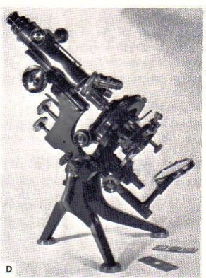
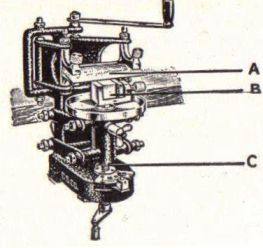
The technology of the microscope advanced: Here are some pictures of 19th century microscopes.

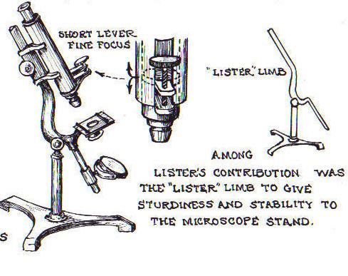
In the next session we'll talk about Joseph Lister, the physician recognized most commonly for establishing the idea of antisepsis in surgery. His father was also named Joseph Lister, Senior, was also a physician, and devised a special combination lens that when put together countered some of the distortions of light that happen in more powerful compound microscopes. This happened in 1830, and made further developments in histology---the just-beginning sub-field of the study of microscopic anatomy---possible.
 
In the ensuing decade the nature of cells was then elaborated; and from this the idea that spontaneous generation was a misleading theory: Only cells give birth to cells. This was also laying the groundwork for germ theory.

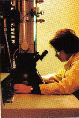
Further developments in microscopy include:


John J. Lister's 1830 microscope, more stable.
 1904 microscope with three possible lenses.

Another requirement for the emergence of histology was the capacity to mount very, very thin sections of tissue so that the overlying elements don't confuse the picture.T

this requires a machine that can slice thinly, a microtome, below left. Along with that is the need to develop ways of fixing the tissues so they don't effectively dissolve or melt---here's where alcohol or formaldehyde was used, so the tissues hold their structure.

In fact, tissues are rather transparent to light, and are not easily visible. Another technology was that of figuring out chemicals that would stain certain parts of a cell and not others, so that their structures could be seen.  Many chemicals were tried and certain ones work.

With bacteria, other stains were found. One kind, the "Gram" stain, colors certain germs such as streptococci and staphylococci blue-black, while others appear red and are called "Gram-negative." This difference became interesting when it appeared that drugs like penicillin affected mainly the former types while other antibiotics sometimes worked with the latter.


 1830 – Joseph Jackson Lister reduces spherical aberration or the "chromatic effect" by showing that several weak lenses used together at certain distances gave good magnification without blurring the image. This was the prototype for the compound microscope.

1830 – Joseph Jackson Lister (the father of the Joseph Lister mentioned in Lecture 2 who introduced antisepsis) reduced the problem of spherical aberration (a distortion of light in strong magnifications) by showing that several weak lenses used together at certain distances gave good magnification without blurring the image.

 1872 – Ernst Abbe, then research director of the Zeiss Optical Works, wrote a mathematical formula called the "Abbe Sine Condition". His formula provided calculations that allowed for the maximum resolution in microscopes possible.
 Other sources:  http://en.wikipedia.org/wiki/Timeline_of_microscope_technologyhttp://www.nationmaster.com/encyclopedia/Timeline-of-microscope-technology

1903 – Richard Zsigmondy developed the ultramicroscope that could study objects below the wavelength of light. He won the Nobel Prize in Chemistry in 1925.

1931 – Ernst Ruska co-invented the electron microscope for which he won the Nobel Prize in Physics in 1986. An electron microscope depends on electrons rather than light to view an object, electrons are speeded up in a vacuum until their wavelength is extremely short, only one hundred-thousandth that of white light. Electron microscopes make it possible to view objects as small as the diameter of an atom.


The electron microscope, which
made it possible to see much smaller:
Here are the smaller fields that modern
microscopy can now see.


The electron microscope allows you to see the insides of cells and identify sub-cellular structures, even within the much smaller bacteria!


This is an electron microscope of a liver cell
This is a diagram of how packed just the edge of the inside of
 an Escheria Coli bacterium can be! (Enlarged 1 million times)

I remember when I studied biology in the 1950s a cell had a more easily stained nucleus and then a vaguely granular "cytoplasm," and most science textbooks didn't have anything about the many components we know know are basic to the cell and operate in that seemingly featureless gray "cytoplasm" area. Our knowledge of sub-cellular structure has grown exponentially since the 1940s.

1932 – Frits Zernike invented the phase-contrast microscope that allowed for the study of colorless and transparent biological materials for which he won the Nobel Prize in Physics in 1953.


1981 – Gerd Binnig and Heinrich Rohrer invented the scanning tunneling microscope that gives three-dimensional images of objects down to the atomic level. Binnig and Rohrer won the Nobel Prize in Physics in 1986. The powerful scanning tunneling mi

Other Technologies

Advances in treatment often awaits the development of the kinds of tools needed to pursue a goal. For example, the relatively recent field of echnology makes a difference. For example, in the past, if a finger or some other part of the body was cut off, efforts at re-implantation often failed, because what was needed was the re-connection ("re-anastomosis") of blood vessels and nerves that were quite small. Our needles and equipment was still to large and clumsy for the job. However, in the mid-20th century, it became technically feasable to make much smaller sutures and needles. Here's a picture of a needle and suture used in microsurgery, placed next to a sesame seed (very enlarged in the picture)! This is the kind of tool needed to put tiny blood vessels back. This surgery also had to be done under very powerful magnifying glasses, mixed with binocular vision for estimating depth.


This illustrates the point that until they figure out how to make certain things it's not easy to figure out how to use them. In other words,  it's not only that necessity is the mother of invention, but sometimes—not infrequently, in fact—invention, or discovery, ends up waking people up to possibilities they hadn’t imagined! We’re seeing that a lot in the computer world, where, given a toy, the game is to find all the things you can do with that toy.
   --- the end for now.